Installation Of New SEM And Sample Preparation Equipment At EMC
The installation of the state-of-the-art FESEM JEOL JSM-IT800 with In-lens Schottky Plus field emission electron gun which can operate at accelerating voltages between 0.01-30 kV is underway. It is replacing JSM 7401F at C256 at Arrhenius lab.
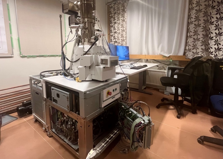
It is equipped with a high-resolution secondary electron detector, a 6-segment annular backscattered electron detector and a new in-lens detector system and the images using different signals can be acquired simultaneously. A motorized annular STEM detector is also present. The instrument can be operated at low vacuum mode in addition to the high vacuum operation. The instrument promises secondary electron image resolution down to 0.6 nm at 15 kV and 1.1 nm at 1 kV.
An SDD detector-based EDS system from JEOL with energy resolution greater than 133 eV to detect B to U as well as a complimentary soft x-ray emission spectrometer (JEOL SXES) with detectable energy range of 50 eV-210 eV enabling detection of elements such as Li are also present.
The new SEM comes with the option to exchange the stage with a cryo-stage operating down to -150°C opening up possibilities for cryo-SEM imaging. To prepare sample for cryo-SEM imaging, a slush freezer and a cryo-workstation are available. A vacuum cryo-transfer system Leica EM VCT 500 is present to facilitate transfer of a frozen specimen from the cryo-work station to the SEM. A cryo-coating unit Leica EM ACE600 is available to freeze fracture and coat the specimen with conducting layer of Pt by sputtering or by e-beam evaporation at cryo-conditions before imaging. VCT 500 can also be used to transfer specimens to SEM under inert conditions.
The SEM will have the option to perform array tomography as well. For automated serial sectioning of the specimen for array tomography, Leica ARTOS 3D is also under installation. In addition to serial sectioning, the ARTOS 3D ultramicrotome can be used to section samples to get thin specimens down to 20 nm for TEM analysis and also block face for SEM and AFM imaging. Sectioning can be carried out at low temperatures for soft specimens using the FC7 cryo-ultramicrotomy option.
A Leica EM ACE600 Sputter/Carbon Thread system is available for coating samples with Pt or C for room temperature SEM analysis. A Cryo Cross-Section Polisher is also under installation which is to prepare cross-sections under cryo and/or inert conditions for SEM imaging.
The new SEM and sample preparation equipment are obtained through the VR infrastructure grant.
Last updated: January 12, 2023
Source: MMK